

| Case Report Online Published: 10 Oct 2024 | ||||||||||||
An interesting case of acute heart failure: peripartum cardiomyopathy Efstathios Koulouris, Evangelos Sdogkos, Aggelos Georgakopoulos, Anila Spahiu, Ioannis Vogiatzis.
| ||||||||||||



Introduction
Peripartum cardiomyopathy (PPCM) is a rare cardiac disorder that develops towards the end of pregnancy or in the first 3 months following delivery (mostly in the month following delivery) and is associated with a high rate of maternal mortality [1]. Although resembling the dilated cardiomyopathy (DCM) phenotype, PPCM is recognized as a distinct entity, different from preexisting cardiomyopathy that is worsened by pregnancy. Its etiology is unknown, although several theories have been reported about its pathogenesis. The clinical presentation is similar to other forms of congestive heart failure but due to the fact that dyspnea and leg swelling are common symptoms during an uncomplicated pregnancy, a high clinical suspicion is needed for the diagnosis [2,3]. Echocardiography is also crucial for the diagnosis and treatment. The management of PPCM is primarily supportive, with the added limitation of the potentially harmful effects of guidelinedirected medical therapy (GDMT) during pregnancy and lactation. While good most of the time, the prognosis in some cases is poor and even cardiac transplantation may be needed [3].
Case Presentation
A 29-yearold woman presented to the emergency department of our hospital with a 7-day history of dyspnea, orthopnea, and bilateral peripheral edema. She had undergone a Csection 10 days before and this was her second childbirth; her first childbirth was uneventful. She was a smoker and her cardiovascular screening while pregnant revealed no abnormalities. Her medical history included gastritis for which she was in pharmaceutical treatment with omeprazole 40 mg once per day. The physical examination, upon arrival, revealed signs of acute heart failure; bilateral crackles on the lower pulmonary fields, an audible S3 sound, and a systolic murmur were heard and bilateral pitting leg edema was observed. Her blood pressure was 145/91 mmHg, her oxygen saturation was 87%, her respiratory rate was 23 per minute and she was afebrile. Neurological examination and examination of the abdomen were normal and the incision from her Csection was not infected or bleeding.
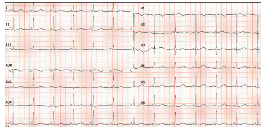
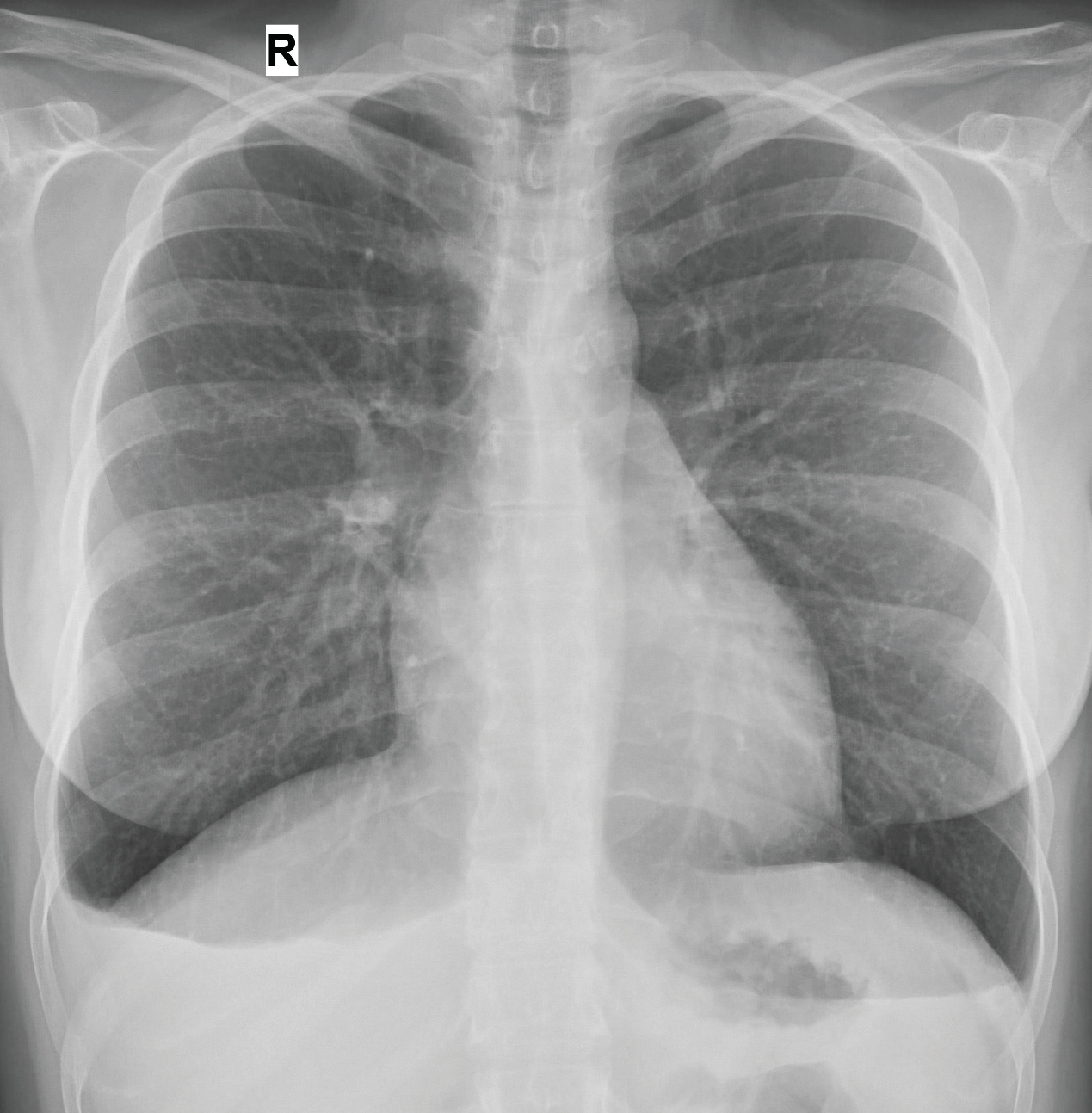
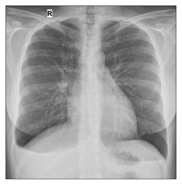
The electrocardiograp (ECG) showed a sinus rhythm with 75 beats per minute with no ischemic abnormalities or arrhythmias (Fig. 1), while her chest Xray showed pulmonary venous congestion with mild cardiomegaly and pulmonary effusions bilaterally (Fig. 2). A transthoracic echocardiography (TTE) was performed that revealed impaired systolic function with ejection fraction (EF)=35% and diffuse hypokinesis of the left ventricle (LV) segments, a small pericardial effusion, elevated filling pressures, and moderate mitral regurgitation.
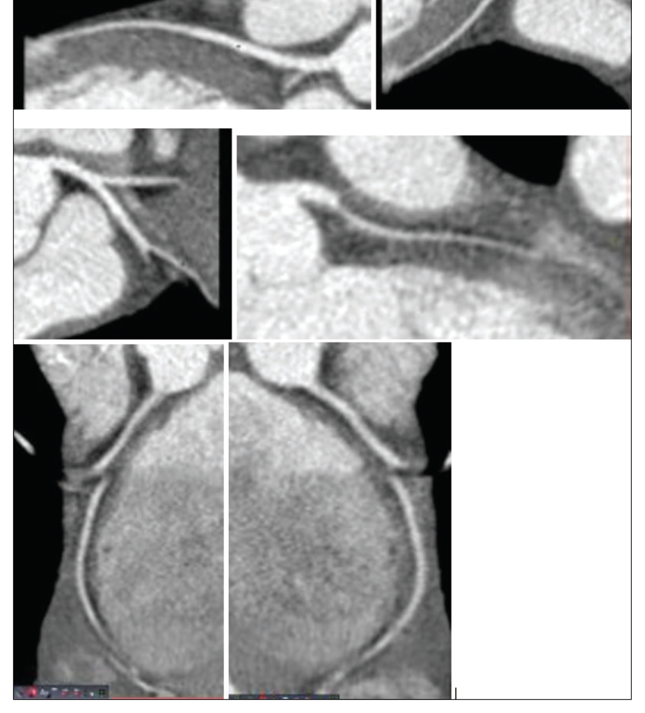
Her blood panel was notable for elevated natriuretic peptide and Ddimer levels, while the enzymes of myocardial damage were within normal range. The patient was admitted to the cardiac intensive care unit and she was placed on 24-hour monitoring. She received treatment with intravenous furosemide, oxygen via a nasal cannula, enalapril, omeprazole, spironolactone per os, and subcutaneous enoxaparin at a prophylactic dose. Computed tomography of the pulmonary arteries was performed because of the hypercoagulable state of the puerperium and no pulmonary embolism was observed. After four days of treatment, the patient was stable with a marked improvement in her clinical status, and beta blockade with metoprolol was added to her medication regimen. No episodes of chest pain, dyspnea, or ventricular arrhythmias were noted and she was no longer in need of oxygenation support. A Coronary computed tomography angiography (CCTA) revealed no stenosis of the coronary arteries and the patient’s calcium score was 0 (Fig. 3). The patient was advised against breastfeeding and she was discharged after an 8-day hospital stay with an urgent referral for a cardiac magnetic resonance imaging (MRI) that took place 15 days after her initial presentation to the hospital. The MRI revealed a small improvement in left ventricular function with EF=45% and areas of late gadolinium enhancement and edema in small areas of the myocardium. Overall, the MRI exam was consistent with the diagnosis of PPCM. The patient remained stable and a full recovery of her cardiac function was observed with TTE three months after her
hospitalization. She was advised not to interrupt her medication regimen for at least 1 year and to contact us in case of a clinical status change. The potential benefits of genetic testing were discussed with her and a close followup was arranged.
Discussion
We report a case of a 29-yearold patient with PPCM who presented with acute heart failure and reduced EF after childbirth. PPCM is an idiopathic cardiomyopathy and is diagnosed after the exclusion of other forms of heart failure such as ischemic heart failure, DCM, or heart failure attributable to myocarditis [4]. PPCM is one of the leading causes of maternal mortality in both developed and less developed countries, with specific rates ranging across different countries and different races [3]. The risk factors associated with PPCM are: age over 30 years, African ancestry, multiparity, multigestation, hypertensive diseases of pregnancy,
anemia, diabetes, obesity, and prolonged tocolysis [5]. Similar to other types of heart failure, PPCM has a highly variable clinical course and patient presentation can consist of mild symptoms to more severe complications like cardiogenic shock. Treatment ought to be individualized and selected according to the opinion of a multidisciplinary team composed of cardiologists, gynecologists, and neonatologists. Even though heart failure GDMT is the cornerstone of PPCM treatment, there are some differences compared to the “typical” heart failure cases. First of all, some heart failure drugs are either contraindicated in pregnancy or lactation or there is insufficient data to ensure safe use. It is obvious that in patients similar to ours who develop PPCM after delivery of the fetus, management is easier since only lactation concerns can hamper our treatment plan (elimination of fetus teratogenicity concerns). It is unclear whether patients with PPCM should stop lactation since there are conflicting data about its effect on prognosis [1]risk factors, clinical presentation, diagnosis and management of PPCM. As shortness of breath, fatigue and leg oedema are common in the peripartum period, a high index of suspicion is required to not miss the diagnosis. Measurement of natriuretic peptides, ECG and echocardiography are recommended to promptly diagnose or exclude heart failure/PPCM. Important differential diagnoses include pulmonary embolism, myocardial infarction, hypertensive heart disease during pregnancy, and preexisting heart disease. A genetic contribution is present in up to 20% of PPCM, in particular titin truncating variant. PPCM is associated with high morbidity and mortality, but also with a high probability of partial and often full recovery. Use of guidelinedirected pharmacological therapy for HFrEF is recommended in all patients respecting contraindications during pregnancy/lactation. The oxidative stressmediated cleavage of the hormone prolactin into a cardiotoxic fragment has been identified as a driver of PPCM pathophysiology. Pharmacological blockade of prolactin release using bromocriptine as a diseasespecific therapy in addition to standard therapy for heart failure treatment has shown promising results in two clinical trials. Thresholds for devices (implantable cardioverterdefibrillators, cardiac resynchronization therapy and implanted longterm ventricular assist devices. A decision should be made after carefully evaluating the possible risks and benefits of continuing lactation.
Besides the wellestablished evidence of the benefit of GDMT, the use of bromocriptine which is a dopamine agonist that suppresses prolactin production, has been associated with more favorable outcomes in some studies [6–8]. However, the flaws of these studies and some conflicting results complicate the decision to administer bromocriptine or not. European guidelines offer an IIb recommendation for bromocriptine in PPCM while in the US its use is not that widespread [9]. Our patient did not have LVEF<35% or right ventricular involvement which are some parameters that have been suggested to enhance the benefit of bromocriptine. Furthermore, we were satisfied with the patient’s response to the treatment and we have no experience with the drug in similar cases, so we decided against administering it to our patient. Genetic testing of patients with PPCM can provide valuable insights regarding risk stratification, treatment choice, treatment duration after LV recovery, and management of future pregnancies [1,3]risk factors, clinical presentation, diagnosis and management of PPCM. As shortness of breath, fatigue and leg oedema are common in the peripartum period, a high index of suspicion is required to not miss the diagnosis. Measurement of natriuretic peptides, ECG and echocardiography are recommended to promptly diagnose or exclude heart failure/PPCM. Important differential diagnoses include pulmonary embolism, myocardial infarction, hypertensive heart disease during pregnancy, and preexisting heart disease. A genetic contribution is present in up to 20% of PPCM, in particular titin truncating variant. PPCM is associated with high morbidity and mortality, but also with a high probability of partial and often full recovery. Use of guidelinedirected pharmacological therapy for HFrEF is recommended in all patients respecting contraindications during pregnancy/lactation. The oxidative stressmediated cleavage of the hormone prolactin into a cardiotoxic fragment has been identified as a driver of PPCM pathophysiology. Pharmacological blockade of prolactin release using bromocriptine as a diseasespecific therapy in addition to standard therapy for heart failure treatment has shown promising results in two clinical trials. Thresholds for devices (implantable cardioverterdefibrillators, cardiac resynchronization therapy and implanted longterm ventricular assist devices.Finally, a factor that may alter our treatment decisions is the common reversibility of the PPCM phenotype. There is a higher threshold for permanent device implantation or heart transplantation in these patients [1]risk factors, clinical presentation, diagnosis and management of PPCM. As shortness of breath, fatigue and leg oedema are common in the peripartum period, a high index of suspicion is required to not miss the diagnosis. Measurement of natriuretic peptides, ECG and echocardiography are recommended to promptly diagnose or exclude heart failure/PPCM. Important differential diagnoses include pulmonary embolism, myocardial infarction, hypertensive heart disease during pregnancy, and preexisting heart disease. A genetic contribution is present in up to 20% of PPCM, in particular titin truncating variant. PPCM is associated with high morbidity and mortality, but also with a high probability of partial and often full recovery. Use of guidelinedirected pharmacological therapy for HFrEF is recommended in all patients respecting contraindications during pregnancy/lactation. The oxidative stressmediated cleavage of the hormone prolactin into a cardiotoxic fragment has been identified as a driver of PPCM pathophysiology. Pharmacological blockade of prolactin release using bromocriptine as a diseasespecific therapy in addition to standard therapy for heart failure treatment has shown promising results in two clinical trials. Thresholds for devices (implantable cardioverterdefibrillators, cardiac resynchronization therapy and implanted longterm ventricular assist devices. Fortunately, our patient did not require any device support.
Conclusion
PPCM is an uncommon but serious cause of heart failure and it usually affects young people who were relatively healthy before the event. It is associated with a high maternal mortality risk, especially in countries with low income. Physicians need to be more aware of the possibility of PPCM and have high clinical suspicion to avoid missing a potentially lifethreatening diagnosis. Specific treatments and strategies mainly regarding the prolactin pathway are currently investigated.
References1. Bauersachs J, König T, van der Meer P, Petrie MC, Hilfiker-Kleiner D, Mbakwem A, et al. Pathophysiology, diagnosis and management of peripartum cardiomyopathy: a position statement from the Heart Failure Association of the European Society of Cardiology Study Group on peripartum cardiomyopathy. Eur J Heart Fail 2019 Jul; 21(7):827–43. 2. Wang M. Peripartum cardiomyopathy: case reports. Perm J 2009; 13(4):42–5. 3. Arany Z. Peripartum cardiomyopathy. N Engl J Med 2024 Jan 11; 390(2):154–64. 4. Bauersachs J, Arrigo M, Hilfiker-Kleiner D, Veltmann C, Coats AJS, Crespo-Leiro MG, et al. Current management of patients with severe acute peripartum cardiomyopathy: practical guidance from the Heart Failure Association of the European Society of Cardiology Study Group on peripartum cardiomyopathy. Eur J Heart Fail 2016 Sep; 18(9):1096–105. 5. Heidenreich PA, Bozkurt B, Aguilar D, Allen LA, Byun JJ, Colvin MM, et al. 2022 AHA/ACC/HFSA guideline for the management of heart failure: a report of the American College of Cardiology/American Heart Association Joint Committee on Clinical Practice Guidelines. Circulation 2022 May 3; 145(18):e895–1032. 6. Hilfiker-Kleiner D, Haghikia A, Berliner D, Vogel-Claussen J, Schwab J, Franke A, et al. Bromocriptine for the treatment of peripartum cardiomyopathy: a multicentre randomized study. Eur Heart J 2017 Sep 14; 38(35):2671–9. 7. Sliwa K, Blauwet L, Tibazarwa K, Libhaber E, Smedema JP, Becker A, et al. Evaluation of bromocriptine in the treatment of acute severe peripartum cardiomyopathy: a proofofconcept pilot study. Circulation 2010 Apr 6; 121(13):1465–73. 8. Tremblay-Gravel M, Marquis-Gravel G, Avram R, Desplantie O, Ducharme A, Bibas L, et al. The effect of bromocriptine on left ventricular functional recovery in peripartum cardiomyopathy: insights from the BRO-HF retrospective cohort study. ESC Heart Fail 2019 Feb; 6(1):27–36. 9. Regitz-Zagrosek V, Roos-Hesselink JW, Bauersachs J, Blomström-Lundqvist C, Cífková R, De Bonis M, et al. 2018 ESC guidelines for the management of cardiovascular diseases during pregnancy. Eur Heart J. 2018 Sep 7; 39(34):3165–241.
| How to Cite this Article |
| Pubmed Style Koulouris E, Sdogkos E, Georgakopoulos A, Spahiu A, Vogiatzis I. An interesting case of acute heart failure: peripartum cardiomyopathy. Case Rep Ser Med. 2024; 1(1): 1-5. doi:10.5455/CRSM.20240907081732 Web Style Koulouris E, Sdogkos E, Georgakopoulos A, Spahiu A, Vogiatzis I. An interesting case of acute heart failure: peripartum cardiomyopathy. https://www.wisdomgale.com/crsm/?mno=302657709 [Access: June 27, 2026]. doi:10.5455/CRSM.20240907081732 AMA (American Medical Association) Style Koulouris E, Sdogkos E, Georgakopoulos A, Spahiu A, Vogiatzis I. An interesting case of acute heart failure: peripartum cardiomyopathy. Case Rep Ser Med. 2024; 1(1): 1-5. doi:10.5455/CRSM.20240907081732 Vancouver/ICMJE Style Koulouris E, Sdogkos E, Georgakopoulos A, Spahiu A, Vogiatzis I. An interesting case of acute heart failure: peripartum cardiomyopathy. Case Rep Ser Med. (2024), [cited June 27, 2026]; 1(1): 1-5. doi:10.5455/CRSM.20240907081732 Harvard Style Koulouris, E., Sdogkos, . E., Georgakopoulos, . A., Spahiu, . A. & Vogiatzis, . I. (2024) An interesting case of acute heart failure: peripartum cardiomyopathy. Case Rep Ser Med, 1 (1), 1-5. doi:10.5455/CRSM.20240907081732 Turabian Style Koulouris, Efstathios, Evangelos Sdogkos, Aggelos Georgakopoulos, Anila Spahiu, and Ioannis Vogiatzis. 2024. An interesting case of acute heart failure: peripartum cardiomyopathy. Case Reports and Series in Medicine, 1 (1), 1-5. doi:10.5455/CRSM.20240907081732 Chicago Style Koulouris, Efstathios, Evangelos Sdogkos, Aggelos Georgakopoulos, Anila Spahiu, and Ioannis Vogiatzis. "An interesting case of acute heart failure: peripartum cardiomyopathy." Case Reports and Series in Medicine 1 (2024), 1-5. doi:10.5455/CRSM.20240907081732 MLA (The Modern Language Association) Style Koulouris, Efstathios, Evangelos Sdogkos, Aggelos Georgakopoulos, Anila Spahiu, and Ioannis Vogiatzis. "An interesting case of acute heart failure: peripartum cardiomyopathy." Case Reports and Series in Medicine 1.1 (2024), 1-5. Print. doi:10.5455/CRSM.20240907081732 APA (American Psychological Association) Style Koulouris, E., Sdogkos, . E., Georgakopoulos, . A., Spahiu, . A. & Vogiatzis, . I. (2024) An interesting case of acute heart failure: peripartum cardiomyopathy. Case Reports and Series in Medicine, 1 (1), 1-5. doi:10.5455/CRSM.20240907081732 |