

| Case Report Online Published: 15 Oct 2024 | ||||||||||||
Elephantiasis gingivae: A report of a rare clinical presentation Mohammad Kaura Abubakar, Mujtaba Bala, Anas Tsafe Bawa.
| ||||||||||||



Introduction
Elephantiasis gingivae otherwise known as hereditary gingival fibromatosis (HGF), idiopathic gingival fibromatosis, congenital hypertrophy of gingiva, congenital macrogingivae, fibromatosis gingivae, and hypertrophic gingiva is a very rare clinical condition characterized by localized or diffuse enlargement of the gingiva [1,2]. The swelling is usually benign, painless, slow growing, fibrous which may involve marginal gingiva, and attached gingiva and seldom extend into the interdental papilla [3]. This overgrown gingival tissue appears normal in color, nonhemorrhagic, firm, and has a characteristic “pebbled” appearance with accentuation of its stippling, which is pathognomonic [3]. Depending on the time the patient presents from the onset of the disease, the enlarged gingival tissue may partially or completely cover the crowns of teeth resulting in serious functional and esthetic concerns [4]. Some of the functional deficits that may occur include difficulty or inability to masticate, speech impairment due to incompetent lips secondary to the bulk of the enlarged gingival tissues, loss of teeth secondary to generalized aggressive periodontitis as a result of the inability to maintain good oral hygiene, and delay or impediment of teeth eruption [2]. It may be present as syndromic or nonsyndromic and can affect both genders [4].The aim of this study is to report a rare case of elephantiasis gingivae and discuss its clinical presentation and management.
Case Report
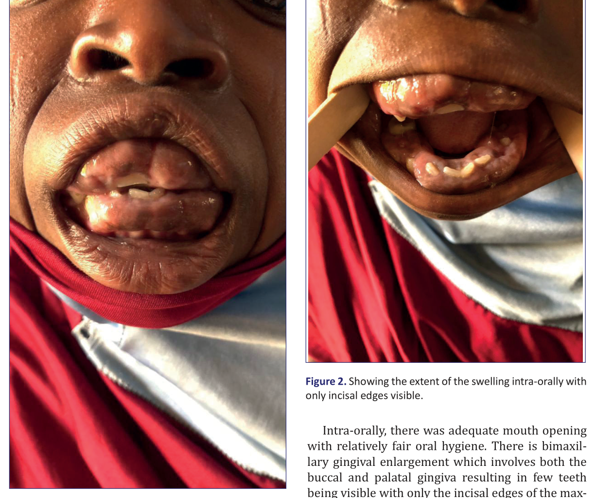
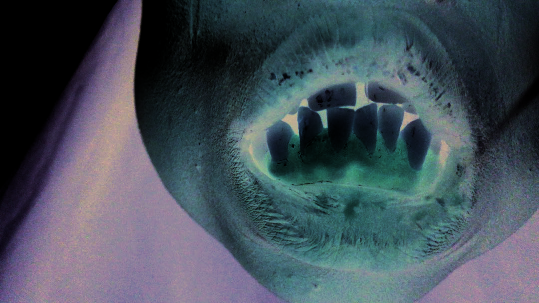
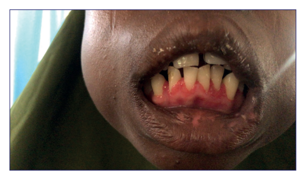
A case a 14-yearold girl presented to the dental and maxillofacial surgery department of Federal Medical Center Gusau with a 4-year history of slow growing, progressive, generalized gingival over growth involving the entire upper and lower gingiva. The patient was totally dependent on a semisolid and liquid diet as the teeth were embedded in the gingiva. Although there was difficulty in taking solid food, the primary patient concern is esthetics. However, there was no significant past medical, dental, family, or postnatal history. On general physical examination, she was found not in obvious distress, acyanosed, anicteric, and not dehydrated, and no signs to suggest a syndrome or any other systemic involvement other than the gingival overgrowth. Extraoral examination revealed facial asymmetry evidenced by a convex facial profile with incompetent lips, normal temporomandibular joint, and nontender nonpalpable lymph nodes, and nil facial hypertrichosis was observed. (Figs. 1 and 2).
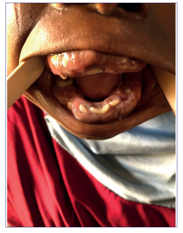
Intraorally, there was adequate mouth opening with relatively fair oral hygiene. There is bimaxillary gingival enlargement which involves both the buccal and palatal gingiva resulting in few teeth being visible with only the incisal edges of the maxillary and mandibular central incisors appearing; also, the cusp tips of the maxillary and mandibular first premolars were visible (Fig. 2). There was the loss of stippling with the gingiva appearing normal in pigmentation, smooth, firm, and fibrotic in consistency. All the teeth were mobile, there was bleeding on probing, and pseudo pockets were present with generalized mobility of teeth. General systemic examination involving cardiovascular, respiratory, gastrointestinal, and musculoskelatal systems revealed normal findings. Hematolotogic, electrolytes urea, and creatinine investigations were within normal range; a Hb of 13 g % was found, and virology was nonreactive. The differential diagnosis may include but not limited to: Idiopathic gingival fibromatosis, druginduced gingival hyperplasia, chronic gingivitis, cherubism, juvenile hyaline fibromatosis, fibroma, pyogenic granuloma, peripheral giant cell granuloma, systemic diseases such as sarcoidosis,


Chron’s disease, and primary gingival tuberculosis. However, detailed history and clinical examination have helped us to narrow down the numerous differential diagnosis to arrive at the provisional diagnosis of Idiopathic gingival fibromatosis. The patient was planned for excisional biopsy (gingivectomy) under general anesthesia having obtained informed consent for the procedure from the parents and the patient assented to it. Consent was also obtained to publish the patient’s photograph. Ethical approval was obtained from the research and ethics committee of the Federal Medical Centre Gusau with reference number FMC/2024/985/009.
Surgical procedure
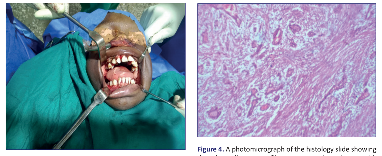
Having observed the routine universal precautions including cleaning and draping of the patient, 2% lignocaine with 1: 200,000 adrenaline was injected around the planned excision intraorally to aid in hemostasis. A waiting time of about 8 minutes was given before a horizontal incision was given at the junction of the fibrotic tissue with the normal mucosa along the mucogingival junction using a size 15 surgical blade beginning with the mandibular facial gingiva followed by the lingual gingiva, and then maxillary facial gingiva and finally the maxillary palatal gingiva. A periosteal elevator was used to tease the incised gingiva away from the teeth and oral mucosa after making another horizontal incision along the gingival margin but sparing it. The rough gingival surface was curetted to remove the remnant fibrotic and inflamed tissues (Fig. 3).Hemostasis was achieved and the patient was placed on postoperative antibiotics and analgesics.
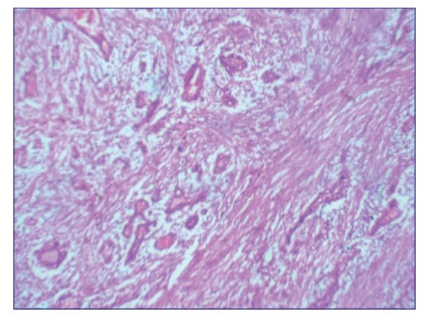
The healing was uneventful in the postoperative period. The final diagnosis was made via histopathological examination of the excised tissue which revealed densely collagenous fibrous connective tissue with haphazard fully differentiated spindleshaped fibrosblasts. The overlying surface epithelium is characterized by thin rete ridges going deeply into the underlying connective tissues. Hence, a final diagnosis of Idiopathic gingival fibromatosis was reached (Fig. 4). Onemonth postoperative followup revealed a satisfactory healing process evidenced by a reduction in teeth mobility, reduction in periodontal pocket depth as well as lack of visible recurrence of the gingival overgrowth (Fig. 5).
Discussion
The index case presented here exhibits classical symptoms of Idiopathic gingival fibromatosis which


are gingival hyperplasia and swelling. There was no prior family history of similar complaints, systemic illnesses, or other potential factors that may induce the occurrence of gingival swellings that may mimic Idiopathic gingival fibromatosis such as some antihypertensive drugs, antidepressants, and immunosuppressants. Idiopathic gingival fibromatosis presents as slowly progressive swelling of the gingiva which cannot be attributed to the irritation caused by plaque secondary to poor oral hygiene [5].As an idiopathic and sometimes hereditary disease, it will be difficult to highlight the mechanism through which the gingival enlargement comes about. However, quite a number of authors have ascribed this phenomenon to excessive collagen accumulation and fibroblast proliferation within the gingival connective tissue [6]. Several efforts have been made to bring to light the genetic and molecular processes involved in the gingival enlargement of GF since 1856 when it was first reported. However, the pathogenic mechanism of the disease entity still remains obscured [6].The commonest form of genetic gingival fibromatosis is HGF which can present in either localized or generalized overgrowth of gingival tissue. In this group, there are two genes that have been for a very long time isolated and attributed to their causation when mutated which are: (SOS1 and REST) in an autosomal dominant pattern of inheritance although there are few reports of an autosomal recessive pattern of inheritance in which case history of consanguinity is almost always present [7]. However, quite recently another novel gene (ZNF862) was found to be related to HGF by Juan et al [8] where they discovered a heterozygous missence mutation (c.2812G > A) in the zinc finger protein 862 gene. Their study supports the biological role of ZNF862 gene for amplifying the profibrotic factors, especially COL1A1 synthesis and ultimately resulting in gingival overgrowth [8]. It was pointed out that the presence of teeth seemed to be a prerequisite for the development of HGF to commence because the gingival enlargement was most severe during the eruptive phase of both deciduous and succedaneous teeth and the condition usually disappears or recedes with the loss of the teeth [9].As a rare condition, characterized by a benign, nonhemorrhagic, localized, or generalized fibrous enlargement of gingiva with slow progression, gingival fibromatosis could be complicated by epilepsy, hypertrichosis, and mental retardation; it can as well develop in relation to syndromes such as hyaline fibromatosis syndrome, Cowden’s syndrome, Ramon syndrome, Zimmerman-Laband syndrome, Cross syndrome, Jones syndrome, Costello syndrome, Ectroamelia syndrome and Rutherford syndrome [10,11]. However, in this case, no such syndrome or any other type was observed. It is generally agreed that the tremendous expansion of gingival tissue observed in GF was mostly secondary to an imbalance between the extracellular matrix (ECM) formation and degradation whereby the collagen formation outweighs its degradation. As a result, this ECM which is mainly made up of collagen will excessively accumulate resulting in gingival fibromatosis. Studies demonstrated that fibroblasts from GF produce 30%–50% more collagen than normal gingival fibroblasts and Type I collagen was the major collagen type produced [12,13].The total elimination of gingival fibromatosis may be faced with a lot of challenges due to the high rate of recurrence. As such, the hallmark of treatment should be centered toward the avoidance of recurrence. The relationship of recurrence with poor oral hygiene is faced with two divergent opinions; while some authors opined that it is observed more in patients with poor oral hygiene, others submitted that the recurrence rate has nothing to do with the oral hygiene status of the patients [14,15]. Several researchers have suggested the use of some biological agents in the management of GF such as the chimeric IgG variant which has been proven to have the ability to inhibit extracellular collagen fibril formation [15]. However, the definitive treatment for severe cases is surgical excision using a scalpel to achieve external bevel gingivectomy. Other viable options are electrosurgery and laser, these two techniques offer some advantages over the conventional surgical approach by reducing intraoperative bleeding and postoperative pain [16]. In our case, we applied the conventional surgical approach under general anesthesia due grotesque nature of the swelling in order to get the full cooperation of the patient.
Conclusion
Elephantiasis gingivae is a rare clinical entity that presents with several esthetic and functional concerns. A surgical approach under general anesthesia may be considered when the size and posterior extent of the gingival swelling are enormous to avoid incomplete surgical excision which may contribute to early recurrence.
Limitation
The longterm success of the intervention could not be ascertained as the patient was lost to followup after 1 month.
Ethical approval and declaration of patient consent Ethical approval was obtained from the research and ethics committee of the Federal Medical Centre Gusau with reference number FMC/2024/985/009. The authors certify that appropriate patient consent was obtained. In the consent form, the patient has agreed and consented to the use of her images and other clinical information to be reported in the journal. The patient is aware that their name and initials will not be published and efforts will be made to conceal her identity, but anonymity may not be guaranteed.
Conflict of interest
There is none declared.
Source of funding
Nil.
References1. Dyasanoor S, Upadhyay K, Naik AR. Elephantiasis gingivae: report of a rare case and literature teview. World J Oral Maxillofac Surg 2019; 2(2):1026. 2. Gawron K, Katarzyna ŁB, Jan P, Maria CG. Gingival fibromatosis: clinical, molecular and therapeutic issues. Orphanet J Rare Dis 2016; 11:9–23; doi 10.1186/s13023-016-0395-13. Livada R, Shiloah J. Gummy smile: could it be genetic? Hereditary gingival fibromatosis. J Mich Dent Assoc 2012; 94(12):40–3. 4. Kanjalkar V, Ashtaputre V. Idiopathic gingival fibromatosis with generalized aggressive periodontitis: a rare case report and its management. IOSR J Dent Med Sci 2012; 1(2):39–43.5. Chapple ILC, Mealey BL, Van Dyke TE, Bartold PM, Dommisch H, Eickholz P, et al. Periodontal health and gingival diseases and conditions on an intact and a reduced periodontium: consensus report of workgroup 1 of the 2017 World Workshop on the classification of periodontal and periimplant diseases and conditions. J Periodontol 2018; 89(Suppl 1):S74–84.6. Almiñana-Pastor PJ, Buitrago-Vera PJ, Alpiste-Illueca FM, Catalá-Pizarro M. Hereditary gingival fibromatosis: characteristics and treatment approach. J Clin Exp Dent 2017; 9(4):e599–602. 7. Pego SPB, Coletta RD, Mendes DC, de Faria PR, Melo-Filho MR, AlvesLR, et al. Hereditary gingival fibromatosis: clinical and ultrastructural features of a new family. Med Oral Patol Oral Cir Bucal 2015 Mar 1; 20(2):e150–5.8. Wu J, Chen D, Huang H, Luo N, Chen H, Zhao J, et al. A novel gene ZNF862 causes hereditary gingival fibromatosis. Elife. 2022 Feb 10;11:e66646; doi 10.7554/eLife.666469. Coletta RD, Graner E. Hereditary gingival fibromatosis: a systematic review. J Periodontol 2006; 77:753–64.10. Balaji P, Balaji SM. Gingival fibromatosis with hypertrichosis syndrome: case series of rare syndrome. Indian J Dent Res 2017; 28:457–60. 11. Guglielmi F, Staderini E, Iavarone F, Di Tonno L, Gallenzi P. Zimmermann-Laband-1 syndrome: clinical, histological, and proteomic findings of a 3-yearold patient with hereditary gingival fibromatosis. Biomedicines 2019; 7:E48. 12. Coletta RD, Almeida OP, Ferreira LR, Reynolds MA, Sauk JJ. Increase in expression of Hsp47 and collagen in hereditary gingival fibromatosis is modulated by stress and terminal procollagen Npropeptides. Connect Tissue Res 1999; 40:237–49. 13. Martelli-Junior H, Cotrim P, Graner E, Sauk JJ. Effect of transforming growth factorbeta1, interleukin-6, and interferongamma on the expression of type I collagen, heat shock protein 47, matrix metalloproteinase (MMP)- 1 and MMP-2 by fibroblasts from normal gingiva and hereditary gingival fibromatosis. J Periodontol 2003; 74:296–306. 14. Bansal A, Narang S, Sowmya K, Sehgal N. Treatment and twoyear followup of a patient with hereditary gingival fibromatosis. J Indian Soc Periodontol 2011; 15:406–9.15. Gawron K, Lazarz-Bartyzel K, Lazarz M, Steplewska K, Pyrc K, Potempa J, et al. In vitro testing the potential of a novel chimeric IgG variant for inhibiting collagen fibrils formation in recurrent hereditary gingival fibromatosis: chimeric antibody in a gingival model. J Physiol Pharmacol 2014; 65:585–91. 16. Aboujaoude S, Aoun G. Hereditary gingival fibromatosis: a report of a severe case. Cureus 2022; 14(3):e23280.
| How to Cite this Article |
| Pubmed Style Abubakar MK, Bala M, Bawa AT. Elephantiasis gingivae: A report of a rare clinical presentation. Case Rep Ser Med. 2024; 1(1): 6-10. doi:10.5455/CRSM.20240913081635 Web Style Abubakar MK, Bala M, Bawa AT. Elephantiasis gingivae: A report of a rare clinical presentation. https://www.wisdomgale.com/crsm/?mno=220465 [Access: June 27, 2026]. doi:10.5455/CRSM.20240913081635 AMA (American Medical Association) Style Abubakar MK, Bala M, Bawa AT. Elephantiasis gingivae: A report of a rare clinical presentation. Case Rep Ser Med. 2024; 1(1): 6-10. doi:10.5455/CRSM.20240913081635 Vancouver/ICMJE Style Abubakar MK, Bala M, Bawa AT. Elephantiasis gingivae: A report of a rare clinical presentation. Case Rep Ser Med. (2024), [cited June 27, 2026]; 1(1): 6-10. doi:10.5455/CRSM.20240913081635 Harvard Style Abubakar, M. K., Bala, . M. & Bawa, . A. T. (2024) Elephantiasis gingivae: A report of a rare clinical presentation. Case Rep Ser Med, 1 (1), 6-10. doi:10.5455/CRSM.20240913081635 Turabian Style Abubakar, Mohammad Kaura, Mujtaba Bala, and Anas Tsafe Bawa. 2024. Elephantiasis gingivae: A report of a rare clinical presentation. Case Reports and Series in Medicine, 1 (1), 6-10. doi:10.5455/CRSM.20240913081635 Chicago Style Abubakar, Mohammad Kaura, Mujtaba Bala, and Anas Tsafe Bawa. "Elephantiasis gingivae: A report of a rare clinical presentation." Case Reports and Series in Medicine 1 (2024), 6-10. doi:10.5455/CRSM.20240913081635 MLA (The Modern Language Association) Style Abubakar, Mohammad Kaura, Mujtaba Bala, and Anas Tsafe Bawa. "Elephantiasis gingivae: A report of a rare clinical presentation." Case Reports and Series in Medicine 1.1 (2024), 6-10. Print. doi:10.5455/CRSM.20240913081635 APA (American Psychological Association) Style Abubakar, M. K., Bala, . M. & Bawa, . A. T. (2024) Elephantiasis gingivae: A report of a rare clinical presentation. Case Reports and Series in Medicine, 1 (1), 6-10. doi:10.5455/CRSM.20240913081635 |