

| Case Report Online Published: 28 Oct 2024 | ||||||||||||||||||||||||||||||
Case Rep Ser Med. 2024; 1(1): 17-21 A Rare Case of Verruca Vulgaris Lip and its Surgical Management Naveen Kumar.
| ||||||||||||||||||||||||||||||



Introduction
Verruca vulgaris commonly known as “wart” is normally seen as skin growth [1, 2]. Verruca vulgaris mostly occurs as an asymptomatic soft tissue lesion on the fingers, toes, soles, and dorsal surfaces of hands. Different types of verrucous and papillary lesions which may be either benign or reactive affect the skin as well as the mucosa of the oral cavity. Oral verruca vulgaris also known as viral papilloma has been commonly associated with human papillomavirus (HPV) 6 and 11 [3]. It is a rare entity in the oral cavity. It can occur at any age with equal gender predilection but is commonly found during the third and fifth decades of life. On clinical examination, it is commonly found as a whitecolored soft tissue lesion but occasionally it can also be seen as a pink lesion having a sessile or pedunculated base with solitary or multiple exophytic, verrucous, or papillomatous projections [4].
It is most commonly seen on the palate followed by the lip, tongue, buccal mucosa, and rarely seen on the gingiva. It is commonly caused by autoinoculation from hand lesions. We present a rare case of largesized verruca vulgaris over lower lip mucosa associated with other leukoplakia lesions [1, 4, 5].
Case Presentation
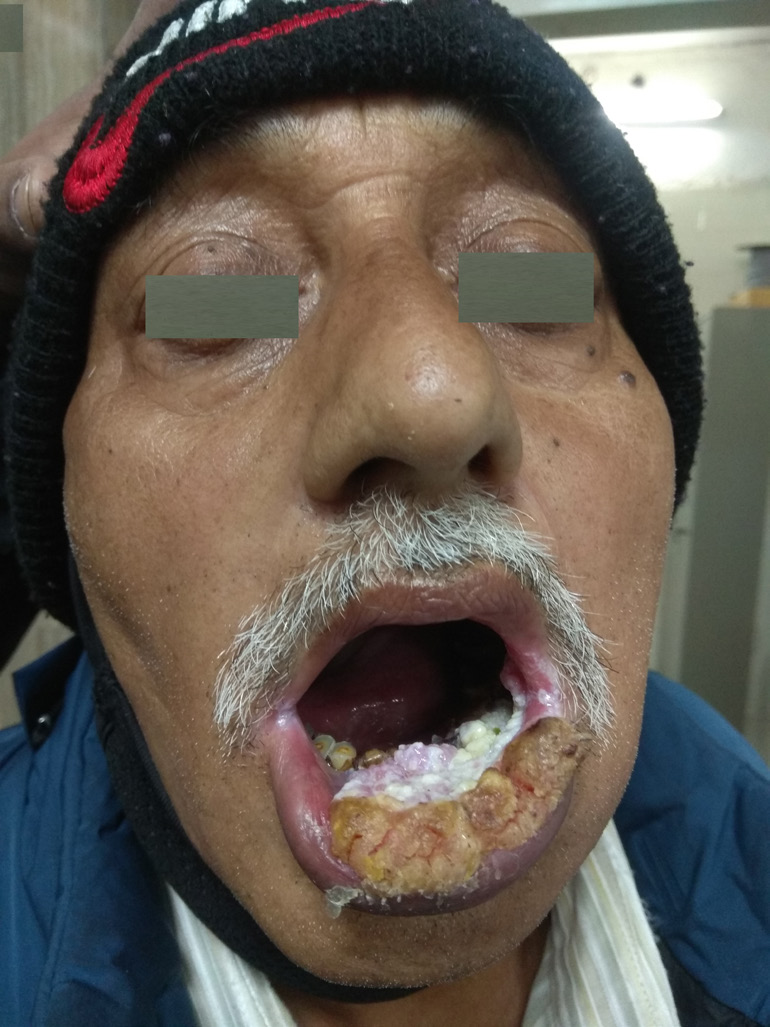
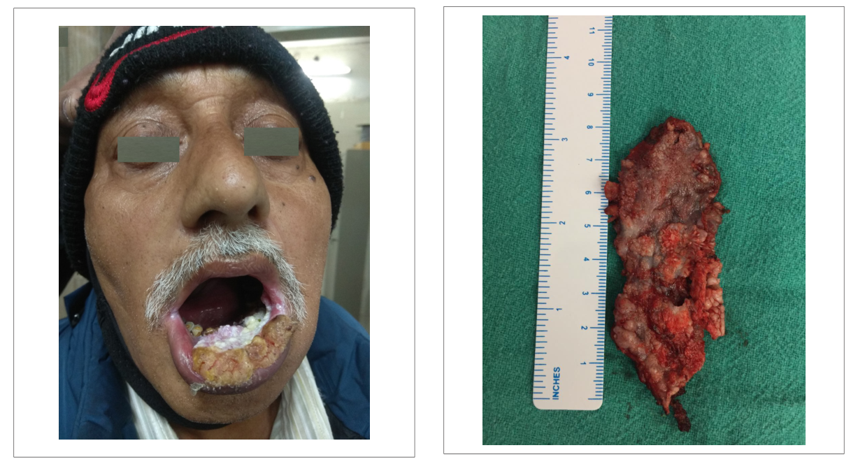
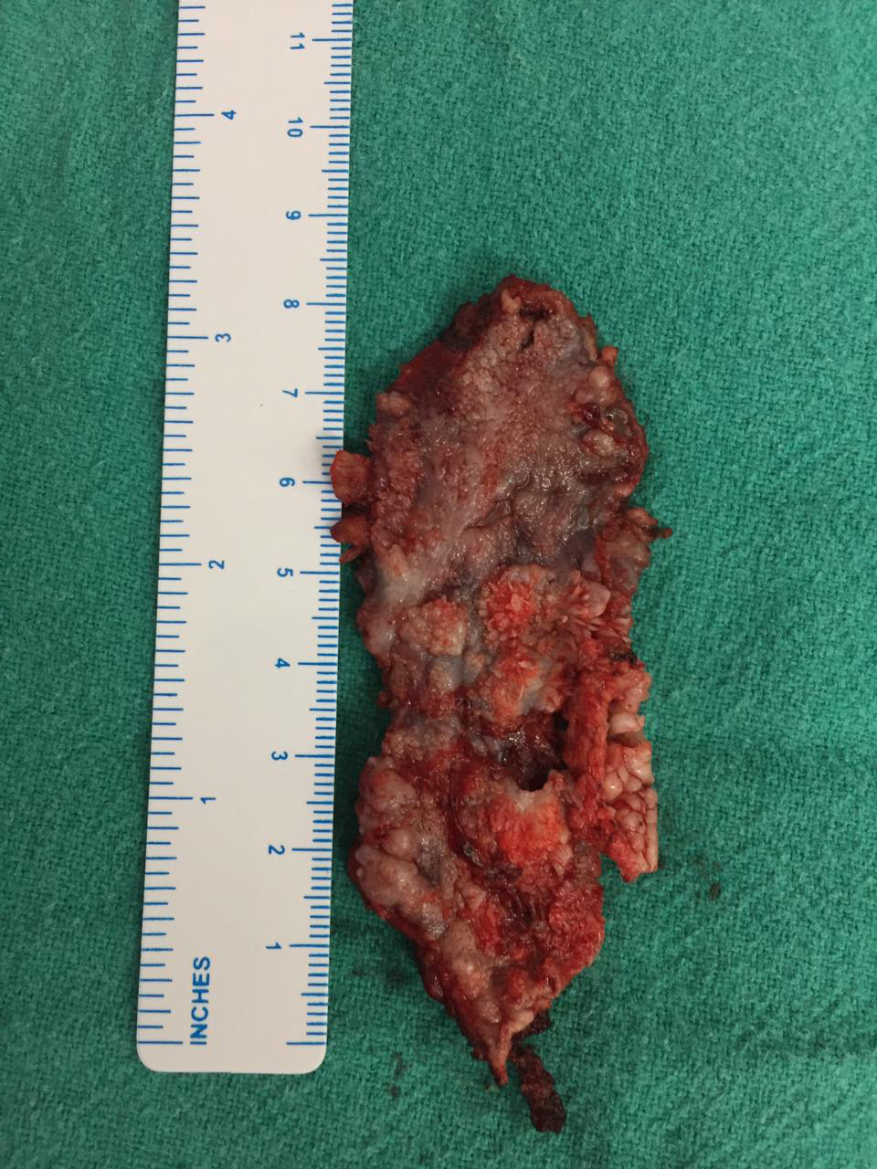
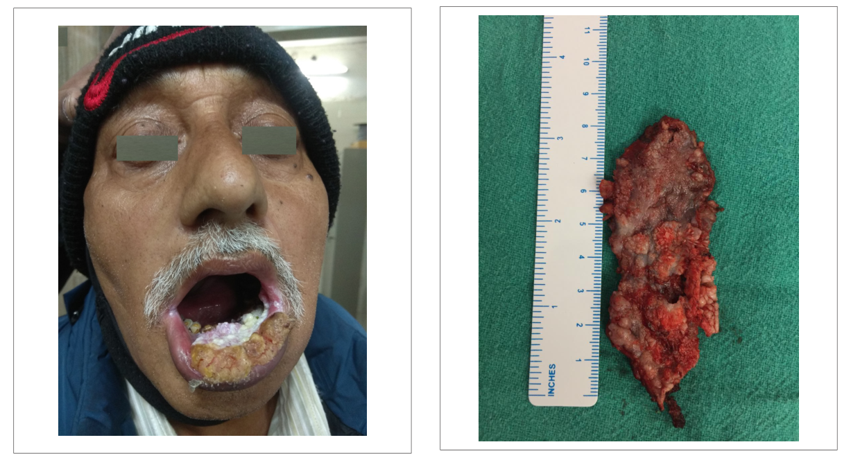
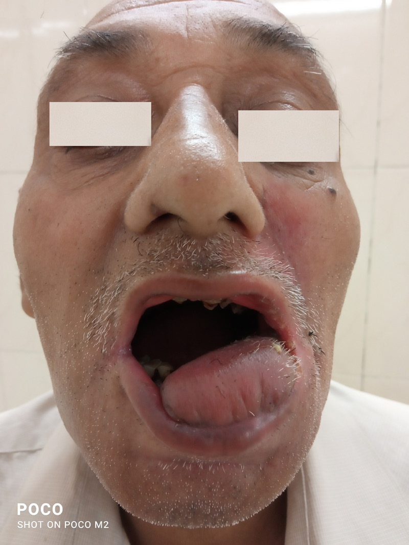
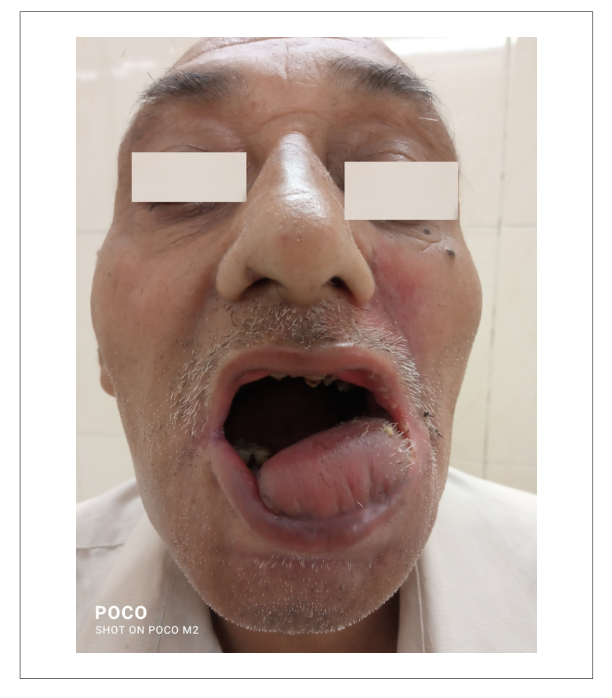
A 60-yearsold male patient (Figure 1) who was a tobacco chewer presented to our department with a complaint of growth on the lower lip for the last 8 months. It was asymptomatic and did not bleed on touch. The patient has been chewing tobacco in the form of pan masala about 10–15 times per day for the last 30 years. Mouth opening was not restricted. On intraoral examination, the oral hygiene was very poor. A single 5 × 3 cm welldefined hyperkeratotic grayishwhite lesion was found with multiple long papillary projections on the mucosal surface of the lower lip involving mostly central and extending to the left commissure. The lesion was nontender and had irregular margins, firm in consistency having a sessile base associated with crusting over the surface along with multiple patches of leukoplakia over the buccal mucosa left side. Neck lymph nodes were not enlarged. No similar lesions were noted elsewhere in the body. A provisional diagnosis of Verrucous leukoplakia or verrucous carcinoma was made. Routine blood investigations with viral markers like HIV, HbsAg test, and multiple incisional biopsies were carried out to rule out other diagnoses. The HIV and HbsAg tests results of the patient were negative. Histopathological examination was suggestive of Verruca Vulgaris which showed stratified squamous epithelium lining the long, fingerlike projections having central connective tissue. The overlying epithelium showed characteristic areas of parakeratosis with irregular hyperchromatic nuclei. Surgical excision of the lesion along with the leukoplakic lesions with safe margins was carried out under general anesthesia. A large buccal mucosal defect of about 7 × 2 cm was created (Figures 2 and 3). Reconstruction was done using an inferiorlybased leftsided Nasolabial flap (Figures 4–6). The postoperative course was uneventful. On followup of 4 months, the patient was completely asymptomatic (Figure 7).
Discussion
The first description about the features of common wart in the past literature was provided by Celsus in 25 AD. In 1848, Tomes coined the term “Gingival wart.” Ciuffo had described the infectious nature of warts in 1907 for the first time [6].
Verruca vulgaris has been described as having
pointed or verruciform surface projections growing along with narrow stalk or sessile base. It’s commonly seen as a firm, exophytic lesion, increasing to the size of up to 5 mm. Such lesions are usually white in appearance due to considerable surface keratin [6].
The association of these lesions has been
described with the Human papillomavirus as an etiological factor. Other risk factors may include tobacco chewing habits or oral submucous fibrosis.
Oral verruca vulgaris is one of the rare lesions that can be presented to a plastic surgeon for the management mimicking the features of exophytic verrucous carcinoma and verruciform xanthoma. Besides these lesions, there are other lesions also which must be kept in mind as differential diagnosis which include verrucous leukoplakia, focal epithelial hyperplasia, exophytic squamous cell carcinoma, squamous papilloma, condyloma acuminatum and keratoacanthoma [6]. Management of these lesions may vary after a definitive diagnosis is established
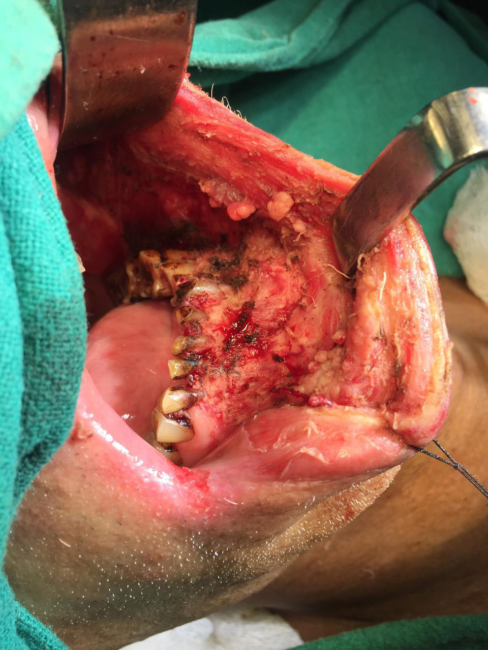
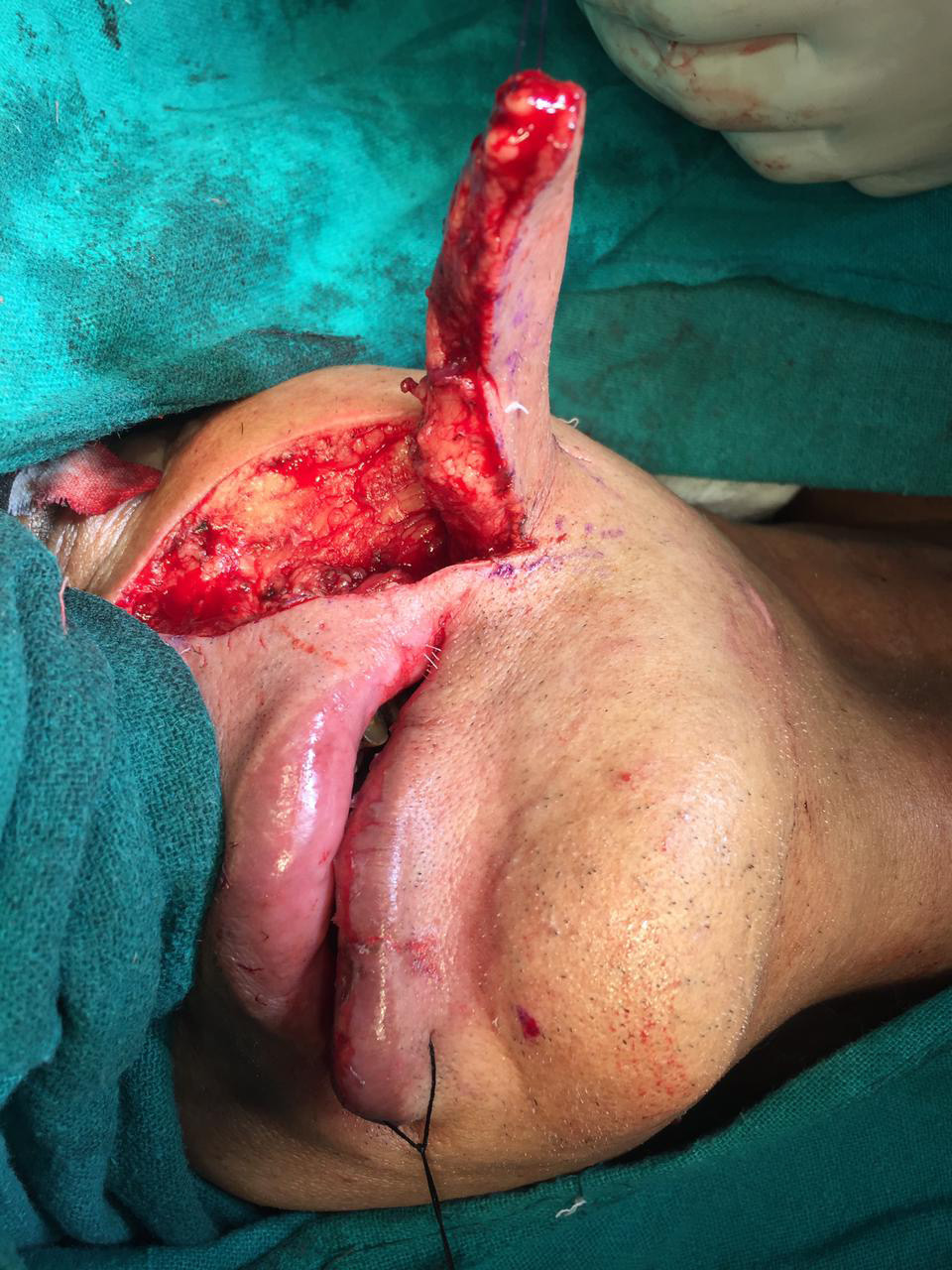
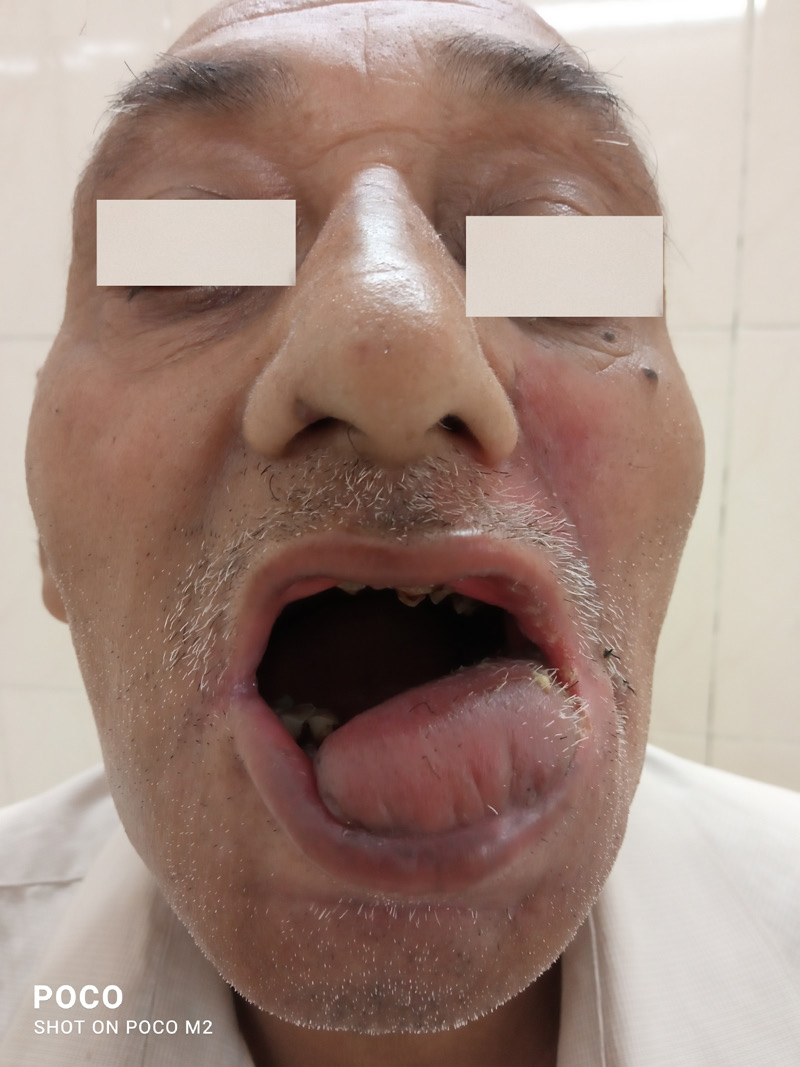
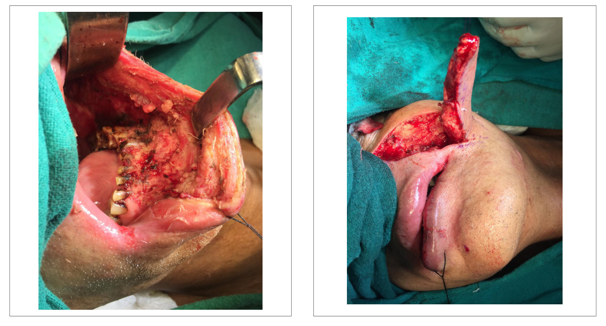
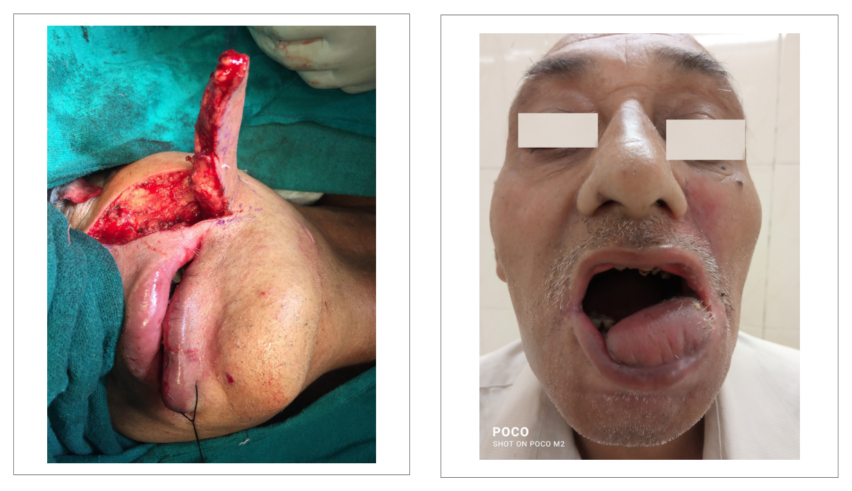
by histological examination. Here we managed the lesion by surgical excision with a safe margin along with the excision of adjacent leukoplakia lesions resulting in a large buccal mucosal defect which we managed with an inferiorly based nasolabial flap.
After a thorough review of the literature, we did not find much of a description of a similar lesion over the lip of size similar to or bigger than the case mentioned above, which was managed with excision and flap cover. Other nonsurgical treatment modalities described include topical salicylic acid, laser, cryotherapy, and retinoids. The cure rates of these nonsurgical modalities vary from 30 to 90% based on individual modalities but these require repeated sessions [7]. However, surgical excision remains the treatment modality of choice. Other than excisional surgery, electrosurgery is also a surgical or physically destructive therapy that can be used for such lesions [7]. Apart from using a local flap for coverage of such defect, a free flap like a radial forearm free flap can also be used. The complications of surgical treatment include scarring, recurrence of lesions, tissue necrosis, or infection [7].
Since these lesions have the propensity to progress into malignant lesion, making it a challenge it a challenge to diagnose and treat at an early stage.
Conclusion
Oral verruca vulgaris is a rare lesion that requires a timely diagnosis and treatment. A surgeon having good clinical acumen with detailed history, clinical examination, and histological features of such lesion will be able to treat the lesion timely and can provide esthetically and functionally better outcomes.
Decalaration
The study is in accordance with the ethical standards of the responsible committee on human experimentation (institutional or regional) and with the helsinki declaration of 1975, as revised in 2000.
Ethics Approval and Consent to Participate
Duly informed consent was obtained in all the cases.The study was approved by the institutional ethic committee.
Contribution
Study design, data collection and analysis, writing of the manuscript.
Conflict of interests
The authors declare that they have no competing interests.
Consent for Publication
Consent to publish has been obtained.
Data availability
Data will be made available upon request at any stage.
Funding source
None.
Acknowledgements
Not applicable.
References1. Swetha P, Supriya NA, Kumar GR. Characterization of different verrucous mucosal lesions. Indian J Dent Res 2013 Sep 1; 24(5):642.
2. Joshi M, Shah A, Vishnoi S. Oral verruca vulgaris: report of two rare cases and review. Natl J Integr Res Med 2013 Jul–Aug; 4(4):145–8.
3. Major T, Szarka K, Sziklai I, Gergely L, Czeglédy J. The characteristics of human papillomavirus DNA in head and neck cancers and papillomas. J Clin Pathol 2005;58:51–5.
4. Sarabadani J, Ghanbariha M, Khajehahmadi S, Nehighalehno M. Consistency rates of clinical and histopathologic diagnoses of oral soft tissue exophytic lesions. J Dent Res Dent Clin Dent Prospects 2009; 3(3):86.
5. Desai VD, Sharma R, Bailoor DN. Extensive mucocutaneous verruca vulgaris in a nonimmunocompromised patient. Int J Clin Pediatr Dent 2011 Jan; 4(1):65.
6. Jigna S, Sharma S. Oral verruca vulgaris – a rare presentation. IOSR JDMS 2017 July; 16(7):74–6.
7. Truong K, Joseph J, Manago B, Wain T. Destructive therapies for cutaneous warts: a review of the evidence. Aust J Gen Pract 2022 Oct; 51(10):799–803. doi: 10.31128/AJGP-01-22-6305
| How to Cite this Article |
| Pubmed Style Naveen Kumar. A Rare Case of Verruca Vulgaris Lip and its Surgical Management. Case Rep Ser Med. 2024; 1(1): 17-21. doi:10.5455/CRSM.20241002015624 Web Style Naveen Kumar. A Rare Case of Verruca Vulgaris Lip and its Surgical Management. https://www.wisdomgale.com/crsm/?mno=222909 [Access: June 26, 2026]. doi:10.5455/CRSM.20241002015624 AMA (American Medical Association) Style Naveen Kumar. A Rare Case of Verruca Vulgaris Lip and its Surgical Management. Case Rep Ser Med. 2024; 1(1): 17-21. doi:10.5455/CRSM.20241002015624 Vancouver/ICMJE Style Naveen Kumar. A Rare Case of Verruca Vulgaris Lip and its Surgical Management. Case Rep Ser Med. (2024), [cited June 26, 2026]; 1(1): 17-21. doi:10.5455/CRSM.20241002015624 Harvard Style Naveen Kumar (2024) A Rare Case of Verruca Vulgaris Lip and its Surgical Management. Case Rep Ser Med, 1 (1), 17-21. doi:10.5455/CRSM.20241002015624 Turabian Style Naveen Kumar. 2024. A Rare Case of Verruca Vulgaris Lip and its Surgical Management. Case Reports and Series in Medicine, 1 (1), 17-21. doi:10.5455/CRSM.20241002015624 Chicago Style Naveen Kumar. "A Rare Case of Verruca Vulgaris Lip and its Surgical Management." Case Reports and Series in Medicine 1 (2024), 17-21. doi:10.5455/CRSM.20241002015624 MLA (The Modern Language Association) Style Naveen Kumar. "A Rare Case of Verruca Vulgaris Lip and its Surgical Management." Case Reports and Series in Medicine 1.1 (2024), 17-21. Print. doi:10.5455/CRSM.20241002015624 APA (American Psychological Association) Style Naveen Kumar (2024) A Rare Case of Verruca Vulgaris Lip and its Surgical Management. Case Reports and Series in Medicine, 1 (1), 17-21. doi:10.5455/CRSM.20241002015624 |